Affinity Chromatography

Enrich a target through a selected ligand interaction — then verify what was captured.
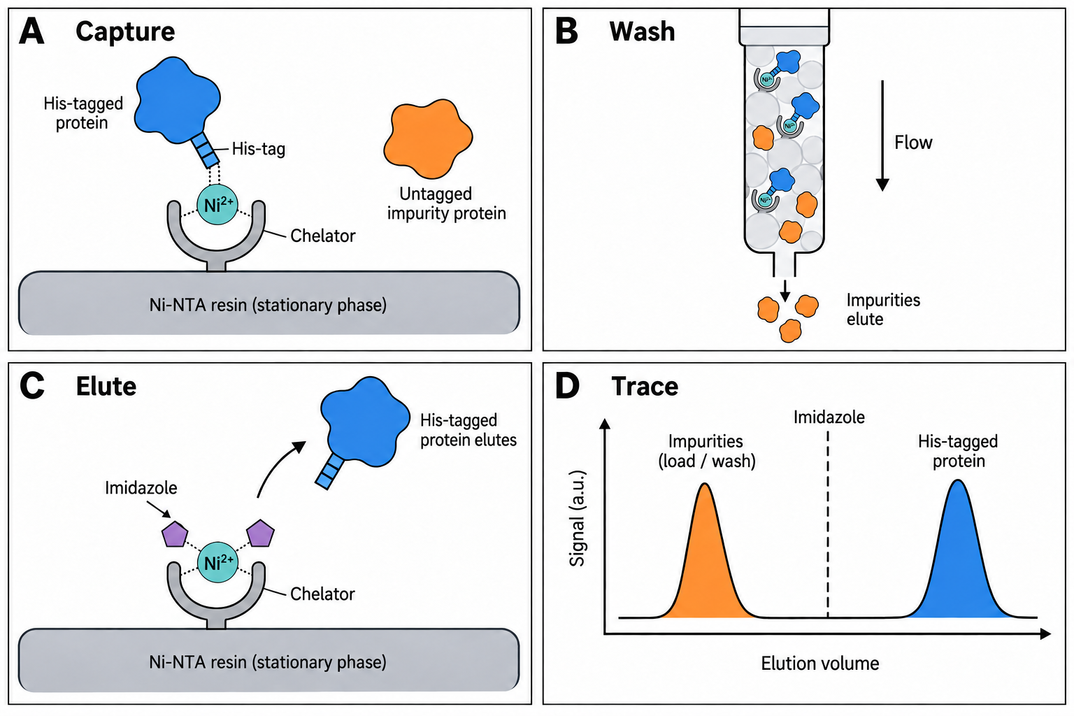
Affinity chromatography, panels A–D. His-tag IMAC example. A: Immobilized nickel coordinates an accessible histidine tag. B: Loading and washing remove less-retained material. C: Imidazole competes with the tag and releases the protein while the metal remains immobilized. The chelator and coordination contacts are symbolic, not a full molecular geometry. D: A retained-protein peak follows elution. Affinity capture enriches a target but does not prove purity or identity; other affinity ligands require different elution conditions.
Affinity chromatography: four-panel overview. His-tag IMAC example. A: Immobilized nickel coordinates an accessible histidine tag. B: Loading and washing remove less-retained material. C: Imidazole competes with the tag and releases the protein while the metal remains immobilized. The chelator and coordination contacts are symbolic, not a full molecular geometry. D: A retained-protein peak follows elution. Affinity capture enriches a target but does not prove purity or identity; other affinity ligands require different elution conditions. Schematics and traces are conceptual, not measured data; a.u. means arbitrary units. Dashed gradient lines, where shown, indicate direction only. Open full-size figure.

Principle

Affinity resins present ligands chosen to bind a tag or protein. They can provide strong enrichment, but ligand specificity is not absolute: host proteins, proteolysis products, leached ligand or metal, and incomplete capture can remain. Choose elution conditions for the particular ligand and sample, then assess identity, contaminants, and function with appropriate orthogonal methods.

His-tag (IMAC)

Nickel or cobalt immobilized through chelating ligands can coordinate a poly-histidine tag. Imidazole is commonly used to tune non-specific binding and elution, but buffer composition, metal choice, and loading conditions need to follow the selected resin manual and be checked on the sample.

  • Screen binding, wash, and elution conditions within the limits stated by the resin manufacturer.
  • Exclude strong chelators such as EDTA unless the resin instructions explicitly allow them.
  • Check reducing-agent compatibility (including DTT) against the selected resin instructions; do not treat it as the same compatibility question as EDTA.
  • Use an orthogonal impurity/identity assay before describing a pool as pure.
Metal coordination in IMAC with IDA and NTA chelatorsThree panels. A: Ni2+ held by IDA through one nitrogen and two oxygens, with three water molecules on the remaining sites. B: Ni2+ held by NTA through one nitrogen and three oxygens, with two histidine side chains of a His-tagged protein on the remaining two sites. C: free imidazole occupies the two sites and the protein is released.A IDA–Ni²⁺ (3 donors)3 positions free (water shown)Ni²⁺H₂ONOH₂OOH₂Omatrix + spacerB NTA–Ni²⁺ + His-tag4 donors; 2 positions bind HisNi²⁺HisNOHisOOmatrix + spacerproteinHis-tagC Elution by imidazolefree Im outcompetes HisNi²⁺ImNOImOOmatrix + spacerproteinreleasedImImImN, O: chelator donor atoms · H₂O: water · His: tag histidine · Im: free imidazole · bold bond: towards viewer; dashed: away
IMAC coordination, schematic (not to scale). Ni²⁺ is shown with six octahedral coordination positions. The tridentate chelator IDA (one N, two carboxylate O) leaves three positions, here occupied by water; the tetradentate NTA (one N, three O) leaves two. On Ni–NTA, two histidine imidazoles of a His-tag can take the two free positions. Excess free imidazole competes for the same positions and displaces the tag; low-pH elution works by protonating histidine instead. Denticities: Zhu et al., Biosensors 2023 (PMC10216662); competitive imidazole and low-pH elution: Cytiva™ HiTrap™ IMAC HP instructions (28404623 AF). Which chelator a given commercial resin uses must be checked in its own documentation.

IMAC: imidazole wash and elution (illustrative model)

Imidazole competes with histidine side chains for the free coordination sites on the immobilized metal. Each protein's bound fraction in one equilibrium stage is modelled as θ = β / (β + (1 + [Im]/K)n), and a wash or elution step as 5 batch stages (θ5 stays bound). The His6 target (β = 2000, n = 2) binds far more strongly than a host protein with one exposed histidine (β = 10, n = 1); K = 10 mM is a scale constant, not a measured dissociation constant.

Fraction still bound versus imidazole concentrationCurves for the His6 target and the host protein fall as imidazole increases; the host curve falls at much lower imidazole. Dashed lines mark the chosen wash (20 mM) and elution (250 mM) concentrations.00.250.50.7510100200300400500Imidazole (mM)Fraction still bound after 5 stagesWashElutionHis6 targethost protein, surface His
Target retained after wash
97.8 %
Host retained after wash
26.9 %
Target released at elution
75 %
Host released at elution
26.9 %
Target share of eluate*
73.6 %

Raising wash imidazole removes more host protein but also strips some target: stringency trades purity against yield. *Equal loaded amounts assumed; real lysates carry far more host protein, and this share is not a purity measurement.

Illustrative relative parameters, not a recipe; purity/yield trade-off and competitive imidazole elution per Cytiva™ HiTrap™ IMAC HP instructions for use (28404623 AF).

GST-tag

Glutathione media bind GST fusion tags. Whether to use competitive elution, cleavage, or a different capture design depends on tag accessibility, target stability, and the downstream use of the material.

Protein A / Protein G (antibodies)

Protein A and Protein G bind many IgG Fc variants, with species/subclass and ligand differences that must be checked in the selected medium’s binding table. Low-pH elution can be effective but may affect a particular antibody; establish recovery and functional integrity for the actual product.

Protein A or G binding the Fc region of an antibodyLeft: an IgG antibody bound by its Fc stem to a Protein A or G ligand on the matrix at neutral binding conditions. Right: after lowering the pH, the antibody has left the ligand.Binding (near-neutral pH)Elution (low pH)matrixProtein A/GmatrixProtein A/GFcFabFabIgG releasedLow pH may affect a particular antibody; establish recovery and function for the actual product.
Protein A or Protein G ligand, coupled to the matrix, binds the Fc region of IgG (schematic, not to scale). Fc binding is the main interaction, but Protein A also binds some Fab variants and Protein G has a low-affinity Fab site. Lowering the pH weakens the interaction and releases the antibody; how low, and whether the antibody tolerates it, is product-specific. Protein A has five Fc-binding domains and one coupled Protein A can bind at least two IgG; species and subclass binding differ between Protein A and G (Cytiva™ Affinity Chromatography Vol. 1: Antibodies, 18103746 AF).

Sources and scope

Wingfield, recombinant-protein purification overview (secondary review, accessed 2026-09-25) supports affinity tags as capture tools, not a guaranteed purity outcome. For operating limits and compatibility, use the exact resin manual; the Cytiva™ handbook index was accessed 2026-09-25. This page intentionally gives no universal buffer recipe.

Try it on a trace

Open a two-column chromatogram in the workbench, or load the synthetic example. No account needed.

Open the workbench

Send feedback

What is it about?

At least 5 characters.0 / 4000

So we can reply. Leave it empty to stay anonymous.

Page: /learn/techniques/affinity